How does one view the brain of an animal that has been dead for over 240 million years? I was fortunate to work on a project that did just that. Under the supervision of Dr. Tim Rowe I worked on the fossilized skull of Thrinaxadon. After taking a 3D X-ray CT scan of the skull I used image processing to enhance structures of the fossilized skull. We used X-rays because this technology was non-destructive and the fossil was irreplaceable. I digitally removed the fossilized skull bones leaving the matrix. The matrix is not an action movie, in this case it is the fossilized stuff that fills in the empty cavities of the skull after the animal dies. The matrix in the braincase of the skull gives a good representation of what the brain looked like when the animal was alive. The process to create a 3D representation of the brain is called making a digital endocast. At the time I was asked if I could remove the fossilized skull and just have the matrix representing the brain. Doing similar digital removals on non fossils I said I could and did. I not only created a 3D model of the brain but also converted the model to a STL format. The STL format allowed anyone with a 3D printer to printout the brain. The Thrinaxadon brain in STL format was published in "Thrinaxadon: Digital Atlas of the Skull" CD-ROM (Second Edition). This 3D model of the skull from a fossilized specimen was the 1st of its kind. Below is an image of the digital 3D Thrinaxadon brain.
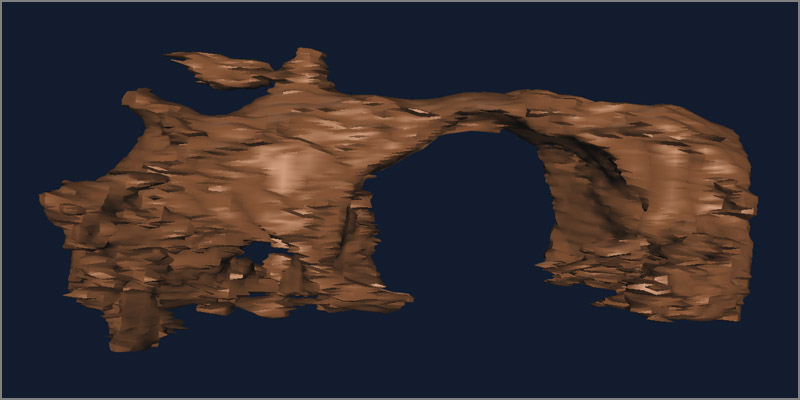
Thrinaxadon liorhinus digital endocast.
The region to the right is not part of the brain, it is the nasal passages in the snout. The highest point in this image is a volcano looking structure,
this is a passage to the Parietal eye on top of the skull. The section to the left of the passage is a gap in the cranial crest that looks like it is part
of the Parietal eye but is not. The back of the brain starts on the left.